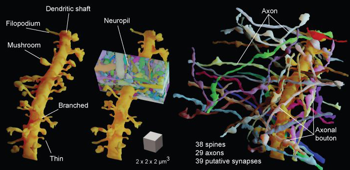
Researchers at the Institute of Science and Technology Austria have developed a brain imaging technique called Live Information Optimized Nanoscopy Enabling Saturated Segmentation (LIONESS). The method lets the researchers create high-resolution 3D images of living brain tissue that reveal its cellular complexity, and even how it changes over time, allowing for monitoring of neural plasticity.
The technique relies on deep learning approaches that help to refine the image quality and also to distinguish cellular structures within this dense and highly complex tissue. The approach can provide unprecedented detail of live tissue without causing damage because it collects as little information as possible, and then uses deep learning to fill in missing information. Without this step, the amount of light required to image in this much detail would fry the living tissue.
The brain is a hugely complex organ, containing approximately 86 billion neurons. These cells are arranged in sophisticated networks and their interconnectivity and activity is also plastic, evolving over time. To date, it has been very difficult to capture this complexity in high-resolution images, particularly without damaging or otherwise compromising the tissue, making it impossible to create such images of living tissue over time.
Previous approaches used to image brain tissue include electron microscopy, which can capture high-resolution images, but requires tissues to be physically sectioned for 3D imaging, which is somewhat of a no-no for living brains. Light microscopy techniques can provide some three-dimensional imaging, but typically with poor resolution.
A more recent approach is called Super-resolution Shadow Imaging, which in combination with a method called Stimulated Emission Depletion can provide high-resolution images that begin to convey the cellular complexity of the brain, but it is not suitable for living tissues, as the amount of light required would still damage the living tissue.
This new approach is designed to provide high-resolution 3D images that convey cellular complexity without cooking the tissue. It uses just enough light to capture the minimum required information for a deep learning algorithm to provide the missing detail. The results are spectacular.
“With LIONESS, for the first time, it is possible to get a comprehensive, dense reconstruction of living brain tissue. By imaging the tissue multiple times, LIONESS allows us to observe and measure the dynamic cellular biology in the brain take its course,” said Philipp Velicky, a researcher involved in the study. “The output is a reconstructed image of the cellular arrangements in three dimensions, with time making up the fourth dimension, as the sample can be imaged over minutes, hours, or days.”
Study in journal Nature Methods: Dense 4D nanoscale reconstruction of living brain tissue
Via: Institute of Science and Technology Austria

